Monday, October 27, 2008

β-blocker drug (Antiadrenegic drug)treatment for IHD

By:Dr.Zharif

β-blocker drug (Antiadrenegic drug)treatment for IHD

Purpose of treatment :

1.Improve quality of life

2.Improve outcome of life


β-blocker drug (Antiadrenegic drug)

General Mechanism of action

block the action of endogenous catecholamines (epinephrine (adrenaline) and norepinephrine (noradrenaline) in particular),

on β-adrenergic receptors, part of the sympathetic nervous system which mediates the fight or flight response.

Action of β adregenic receptor (part of sympathethic nerves system)

-three known types of beta receptor, designated β1, β2 and β3.

a) β1-Adrenergic receptors are located mainly in the heart and in the kidneys.

b) β2-Adrenergic receptors are located mainly in the lungs, gastrointestinal tract, liver, uterus, vascular smooth muscle, and skeletal muscle.

c) β3-receptors are located in fat cells

Respiratory system(β2 agonists receptor)

1.bronchodilation

2. Increase Respiratory Secretions

Cardiovascular system

Heart(β1 agonists receptor)

1.Increase heart rate

2.Increase AV conduction

3.Increase BP -rise in systolic and diastolic-NA

-rise in systolic and fall diastolic-Iso

Blood vessel(β2 agonists receptor)

1.Vasodilation

Eye(β2 agonists receptor)

1.Increase secretory activity of ciliary epithelia(caution to patient with glaucoma)

2.Increase Intraocular Pressure

GIT(not clinical significant)

1.Reduced peristalsis

2.Sphincter constricted

Bladder

1.Relaxed detrussor muscle

Uterus

1.Decrease contraction of uterus

Metabolic

1.Increase glycogenolysis- hyperglycaemia,hypelactidemia(β2)

2.Decrease insulin

3.Increase lipolysis

4.Increase lipid profile in plasma

5.Transient hyperkalemia followed by hypokalemia

CNS

Not produce any effect on CNS because poorly penetration in brain (hydrophilic)

Lipophilic drugs

Lipophilic drugs (metoprolol, propranolol, timolol) are

rapidly and completely absorbed from the gastr

ointestinal

tract but are extensively metabolised in the gut wall

and in the liver (first pass effect), so that the

ir oral

bioavailability is low (10–30%). These drugs may accumulate

in patients with reduced hepatic blood flow (i.

e.,

elderly, congestive heart failure, liver cirrhosis). Lipophilic

drugs present short elimination half-lives (1-5 h)

and they easily enter the central nervous sys

tem (CNS),

which may account for a greater incidence of

central

side-effects.

Hydrophilic drugs

Hydrophilic drugs (atenolol, esmolol) are absorbed incompletely

from the gastrointestinal tract and are excreted

unchanged or as active metabolites by the kidney.

They have longer half-lives (6–24 h), and do not interact

with other liver-metabolised drugs. They barely cross the

blood–brain barrier. Elimination half-life is increased

when glomerular filtration rate is reduced (i.e., elderly,

renal insufficiency).

Mechanism of action in IHD

The prevention of the cardiotoxic effects of catecholamines plays

The following mechanisms are also considered:

(a) Antihypertensive action.

Associated with a decrease in cardiac

output, inhibition of the release of renin and production

of angiotensin II, blockade of presynaptic a-adrenoceptors

that increase the release of norepinephrine from

sympathetic nerve terminals and decrease of central

vasomotor activity.1–9

(b) Anti-ischaemic action

b-blockers decrease myocardial oxygen demand by reducing

heart rate, cardiac contractility, and systolic

blood pressure.10 In addition, prolongation of diastole

caused by a reduction in heart rate may increase myocardial

perfusion.

(c) Reduction of renin release and

angiotensin II and aldosterone production by blocking of

b1-adrenoceptors on renal juxtaglomerular cells.

(d)Improvement of left ventricular structure and function,

decreasing ventricular size and increasing ejection fraction.

6–8 b-blockers may improve cardiac function because

they:

(i) reduce heart rate, prolong diastolic filling

and coronary diastolic perfusion time,

(ii) decrease myocardial

oxygen demands,

(iii) improve myocardial energetics

by inhibiting catecholamine-induced release

of free fatty acids from adipose tissue,

(iv) upregulate

b-adrenergic receptors

(v) reduce myocardial

oxidative stress.1;11;12

(e) The antiarrhythmic effect, the

result of direct cardiac electrophysiological effects

(reduced heart rate, decreased spontaneous firing of

ectopic pacemakers, slowed conduction and increased

refractory period of AV node),

reduces the sympathetic

drive and myocardial ischaemia, improves baroreflex

function and prevents catecholamine-induced hypokalemia.

Other mechanisms include:

Inhibition of cardiac apoptosis mediated via the activation of the b-adrenergic

pathway,

Inhibition of platelet aggregation,

Reduction of the mechanical stress imposed on t

he plaque,

Preventing plaque rupture

Resensitization of the b-adrenergic pathway and changes in myocardial gene expression,

i.e., an increase in sarcoplasmic reticulum calcium ATPase, mRNA and a-myosin heavy chain mRNA and a decrease in b-myosin heavy chain mRNA levels.15

exhibit antioxidant properties and inhibit vascular smooth

muscle cell proliferation.

Other Indication

· Hypertension

· Angina

· Mitral valve prolapse

· Cardiac arrhythmia

· Congestive heart failure

· Myocardial infarction

· Glaucoma

· Migraine prophylaxis

· Symptomatic control (tachycardia, tremor) in anxiety and hyperthyroidism

· Essential tremor

· Phaeochromocytoma, in conjunction with α-blocker

also been used in the following conditions

· Hypertrophic obstructive cardiomyopathy

· Acute dissecting aortic aneurysm

· Marfan syndrome (chronic treatment with propranolol slows progression of aortic dilation and its complications)

· Prevention of variceal bleeding in portal hypertension

· Possible mitigation of hyperhidrosis

· Social anxiety disorder and other anxiety disorders

Adverse effect

Cardiovascular

1.Reduce heart rate,

2.Extreme bradycardia and AV block.

3.Impaired sinusnode function and AV-node conduction

4.Produce cold extremities and Raynaud’s phenomenon and worsen the symptoms in patients with severe peripheral vascular disease.

5.Coronospasm

Metabolic

Hypoglycaemia -In patients with insulin-dependent type I diabetes nonselective

b-blockers mask and selective b-blocker should therefore be preferred at

least in insulin dependent patients

In one study carvedilol decreased the new onset diabetes in

patients with heart failure.

Pulmonary

Increase in airway resistance and are contraindicated in patients with

asthma or bronchospastic chronic obstructive pulmonary

disease.

Central effects

Central effects (fatigue, headache, sleep disturbances,

insomnia and vivid dreams, depression) are less common

with hydrophilic drugs

In some patients the fatigue may

be related to a decrease in blood flow to skeletal muscles;

in other cases, it may be secondary to a central effect.

Sexual dysfunction

Impotence and loss of libido.

Abrupt discontinuation of b-blockers after chronic

treatment can lead to rebound symptoms (i.e., hypertension,

arrhythmias, exacerbated angina)

This increased risk is related with upregulation of badrenoceptors during chronic treatment

Contraindications

  1. Asthma,
  2. Symptomatic hypotension
  3. Bradycardia
  4. Severe decompensated heart failure
  5. Chronic obstructive lung disease without bronchospastic activity
  6. Peripheral vascular disease are not considered as absolute contraindications and high risk patients may obtain a significant benefit from this therapy.
  7. Patients with heart failure and bradycardia due to sick sinus node or second or third degree AV-block may benefit from pre-treatment with pacemaker in order to tolerate b-blockers, although this approach has, however, not been formally tested.
  8. Diabetes or intermittent lower limb claudication are not absolute contraindications for b-blockers use

I. Non-selective β1, β2 adrenergic antagonists

Carteolol + Low 2.5–20 mg once/twice daily

Nadolol 0 Low 40–320 mg once daily

Penbutolol + Moderate 20–80 mg once/twice daily

Pindolol ++ High 10–40 mg twice daily

Propranolol 0 High + 40–180 mg twice daily

Sotalol 0 Low +

Timolol 0 High 5–40 mg twice daily

II. Selective β1adrenergic antagonists

Acebutolol + Moderate 200–800 mg once/twice daily

Atenolol 0 Low + 25–100 mg once daily

Betaxolol 0 Moderate 5–20 mg once daily

Bisoprolol 0 Moderate 2.5–10 mg once daily

Celiprolol + Moderate 200–600 mg once daily

Esmolol 0 Low Only i.v.

Metoprolol 0 High 50–100 mg once/twice daily

Nevibolol 0 2.5–5 mg once daily

III. α1- and β1-adrenergic antagonists

Bucindolol + Moderate 25–100 mg twice daily

Carvedilol_ 0 Moderate 3.125–50 mg twice daily

Labetalol + Low 200–800 mg twice daily

ISA: Intrinsic Sympathomimetic Activity;

i.v.: Intravenous administration possible; AMI: Acute Myocardial Infarction; CHF: Chronic Heart Failure.

Acute Myocardial Infarction (AMI)

Intravenous dosing of b-blockers

Drug Loading dose Maintenance dose

Atenolol 5 ю 5 mg Oral, 50–100 mg/day

Esmolol 0.5 mg/kg over 1–5 min 0.05–0.3 mg/kg/min

Labetalol 20 mg in 2 min 2–10 mg/min

Metoprolol 2.5–5 mg i.v. bolus over 2 min; up to three doses Oral, 25–100 mg/12 h

Propranolol 0.15 mg/kg 0.10–0.20 mg/kg/min oral, 80–240 mg/day


Practical guidance on using b-adrenergic blockers in heart failure

Who should receive b-blocker therapy

_ All patients with chronic, stable heart failure

_ Without contraindications (symptomatic hypotension or bradicardia, asthma)

What to promise

Treatment is primarily prophylactic against death and new hospitalisations for cardiovascular reasons. Some patients will

experience improvement of symptoms.

When to start

_ No physical evidence of fluid retention (use diuretics accordingly)

_ Start ACE-I first if not contraindicated

_ In stable patients, in the hospital or in outpatient clinics

_ NYHA class IV/severe CHF patients should be referred for specialist advice

_ Review treatment. Avoid verapamil, diltiazem, antiarrhythmics, non-steroidal anti-inflamatory drugs

Beta-blocker

_ Bisoprolol, carvedilol or metoprolol

Dose

_ Start with a low dose

_ Increase dose slowly. Double dose at not less than 2 weekly intervals

_ Aim for target dose (see above) or, if not tolerated, the highest tolerated dose

Starting dose mg Target dose mg

Bisoprolol 1.25 once daily 10 once daily

Carvedilol 3.125 twice daily 25–50 twice daily

Metoprolol CR/XL 12.5–25 once daily 200 once daily

Monitoring

_ Monitor for evidence of heart failure symptoms, fluid retention, hypotension and bradycardia

_ Instruct patients to weigh themselves daily and to increase their diuretic dose if weight increases

Problem solving

_ Reduce/discontinue b-blocker only if other actions were ineffective to control symptoms/secondary effects

_ Always consider the reintroduction and/or uptitration of the b-blocker when the patient becomes stable

_ Seek specialist advice if in doubt.

Symptomatic hypotension (dizziness, light headedness and/or confusion)

_ Reconsider need for nitrates, calcium channel blockers and other vasodilators

_ If no signs/symptoms of congestion consider reducing diuretic dose

Worsening symptoms/signs (increasing dyspnoea, fatigue, oedema, weight gain)

_ Double dose of diuretic or/and ACE-I.

_ Temporarily reduce the dose of b-blockers if increasing diuretic dose does not work

_ Review patient in 1–2 weeks; if not improved seek specialist advice

_ If serious deterioration halve dose of b-blocker

_ Stop b-blocker (rarely necessary; seek specialist advice)

Bradycardia

_ ECG to exclude heart block

_ Consider pacemaker support if severe bradycardia or AV block or sick sinus node early after starting b-blockers

_ Review need, reduce or discontinue other heart rate slowing drugs, e.g., digoxin, amiodarone, diltiazem

_ Reduce dose of b-blocker. Discontinuation rarely necessary

Severe decompensated heart failure, pulmonary oedema, shock

_ Admit patient to hospital

_ Discontinue b-blocker if inotropic support is needed or symptomatic hypotension/bradycardia is observed

_ If inotropic support is needed, levosimendan may be preferred


Reference
1. Cycle clinical farmacology 6th year RSMU
2.Essential of Medical Pharmacology,KD Tripathi,JAYPEE
3.wikipedia
4.Escardio(Europian Society of Cardiology)
Sunday, October 26, 2008

Conducting systems of the Heart

By:Dr.Norzaher
There are four basic components to the heart's conduction system

(1) sinoatrial node (SA node)

(2) inter-nodal fibre bundles

(3) atrioventricular node (AV node)

(4) atrioventricular bundle

The sinoatrial (SA) node is a small mass of specialised cardiac muscle situated in the superior aspect of the right atrium. It lies along the anterolateral margin of this chamber between the orifice of the superior vena cava and the auricle. The specialised cardiac muscle of the SA node is characterised by the property of automatic self-excitation and it initiates each beat of the heart. Therefore, the SA node is often referred to as the pacemaker of the heart.

Since the fibres of the SA node fuse with the surrounding atrial muscle fibres, the action potential generated in the nodal tissue spreads throughout both atria at a rate of approximately 0.3 meter per second and produces atrial contraction. Interspersed among the atrial muscle fibres are several inter nodal fibre bundles which conduct the action potential to the atrioventricular (AV) node with a greater velocity (approximately 1.0 meter per second) than ordinary atrial muscle. The AV node is located in the right atrium near the lower part of the interatrial septurn. Here there is a short delay (approximately 0.1 second) in transmission of the impulse to the ventricles.

This is important because it permits the atria to complete their contraction and empty their blood into the ventricles before the ventricles contract. The delay occurs within the fibres of the AV node itself as well as in special junctional fibres that connect the node with ordinary atrial fibres.

Once the action potential leaves the AV node, it enters specialised muscle fibres called Purkinje fibres. These are grouped into a mass termed the atrioventricular (AV) bundle, or the bundle of His. The Purkinje fibres are very large and conduct the action potential at about six times the velocity of ordinary cardiac muscle (i.e., 1.5 to 4.0 meters per second). Thus the Purkinje fibres permit a very rapid and simultaneous distribution of the impulse throughout the muscular walls of both ventricles.

As the AV bundle leaves the AV node, it descends in the interventricular septurn for a short distance and then divides into two large branches, the right and left bundle branches. Each of these descends along its respective side of the interventricular septum immediately beneath the endocardium and divides into smaller and smaller branches. Terminal Purkinje fibres extend beneath the endocardium and penetrate approximately one-third of the distance into the myocardium. Their endings terminate upon ordinary cardiac muscle within the ventricles, and the impulse proceeds through the ventricular muscle at about 0.3 to 0.5 meters per second. This results in a contraction of the ventricles that proceeds upward from the apex of the heart toward its base. The pathway taken by each action potential generated by the SA node is represented schematically as

The spontaneous generation of an action potential within the SA node initiates a sequence of events known as the cardiac cycle. Each cardiac cycle lasts approximately 0.8 second and spans the interval from the end of one heart contraction to the end of the subsequent heart contraction.

Ordinarily this occurs about 72 times each minute.

The cardiac cycle has two basic components:

(1) a contraction phase (systole) during which blood is ejected from the heart

(2) a relaxation phase (diastole) during which the chambers of the heart are filled with blood.

The spontaneous generation of an action potential within the SA nodal tissue represents the start of the cardiac cycle. This electrical impulse spreads throughout the atrial muscle and leads to contraction of the two atria.

As the atria contract, the AV valves remain open and additional blood is forced into the ventricles from the veins. A large amount of blood has already passed from the atria to the ventricles prior to atrial contraction.

The aortic and pulmonary (pulmonic) semilunar valves remain closed .

After the ventricles have filled (mostly by blood returning from the large veins) and the atria have contracted, the AV valves close as the ventricles begin their contraction.

Ventricular contraction forces blood through the semilunar valves into the aorta and pulmonary trunk.

Next, as the ventricles begin to relax, the aortic and pulmonic semilunar valves close, the AV valves open, and blood flows into the ventricles to begin another cycle.

While the atria are in systole, the ventricles are relaxed (in diastole). The atria relax during ventricular systole and remain in this phase even during a portion of ventricular diastole.

Blood (like any other fluid) tends to flow from a region of high pressure to one of lower pressure.

As each chamber of the heart fills with blood, the pressure increases within it. The blood moves out of the chamber, when the various one-way valves guarding those chambers permit it to do so.

For example, intra-atrial pressure increases as blood from the veins enters them, and this pressure increases further during atrial systole.

As the ventricles contract, the blood is forced in a retrograde fashion against the AV valves, which causes them to bulge inward slightly toward the atria and which also elevates atrial pressure.

In doing so, the AV valves are effectively closed and blood is prevented from regurgitating back into the atria. Near the end of ventricular systole the AV valves are still closed and since the atria are in the process of filling, this too contributes to a rise in intra-atrial pressure.

Even before the atria enter systole, the ventricles are filled with blood to approximately 70% of their capacity. When the atria do finally contract, additional blood enters the ventricles and elevates the intraventricular pressure. As the ventricles contract, blood is forced backward, closing the AV valves, and a sharp rise in ventricular pressure occurs.

Although the ventricles exist as closed chambers for a brief moment, the pressure within them soon exceeds that in the aorta and pulmonary trunk. When this happens the aortic and pulmonic semilunar valves are forced open under pressure and blood rushes out of the ventricles and is driven into these large vessels. Accompanying the opening of the semilunar valves is a rapid decline in intraventricular pressure that continues until the pressure within the ventricles becomes less than that of the atria. When this pressure differential is reached, blood within the atria pushes the AV valves open and begins to fill the ventricles once again.


Composition of the Heart

The heart comprises of three layers, epicardium, myocardium, and endocardium surrounding the inner chambers and is enclosed in a fourth protective layer known as the pericardium.

The image below shows a section through the wall of the heart in the middle of the right ventricle. Note the branched appearance of the cardiac muscle in the myocardium.


The pericardium

Within the mediastinum, the heart and origins of the great blood vessels are enclosed in a loose fitting sac termed the pericardium. This protective sac is composed of two layers separated by a space called the pericardial cavity. The outer layer is termed the parietal pericardium, and the inner layer is called the visceral pericardium.

The parietal pericardium consists of an outer layer of thick, fibrous connective tissue and an inner serous layer. The serous layer, consisting largely of mesothelium together with a small amount of connective tissue, forms a simple squamous epithelium and secretes a small amount of fluid. Normally the total volume of this fluid is only about 25 to 35 ml.

This fluid layer lubricates the surfaces to allow friction free movement of the heart within the pericardium during it's muscular contractions.

The fibrous layer of the parietal pericardium is attached to the diaphragm and fuses with the outer wall of the great blood vessels entering and leaving the heart. Thus, the parietal pericardium forms a strong protective sac for the heart and serves also to anchor it within the mediastinum.

The visceral pericardium is also known as the epicardium and as such comprises the outermost layer of the heart proper.

The epicardium (or visceral pericardium) forms the outer covering of the heart and has an external layer of flat mesothelial cells. These cells lie on a stroma of fibrocollagenous support tissue, which contains elastic fibres, as well as the large arteries supplying blood to the heart wall, and the larger venous tributaries carrying blood from the heart wall. The large arteries (coronary arteries) and veins are surrounded by adipose tissue, which expands the epicardium.

The image on the left is a diagrammatic representation of a section through the left and right ventricles as viewed from above.

Notice the difference in thickness between the wall of the left ventricle and that of the right ventricle. Also note their different shapes.

Atrial myocardial fibres are not only smaller than those of the ventricles but also contain small neuroendocrine granules, which are usually sparse and located close to the nucleus; they are most numerous in the right atrium. These granules secrete atrial natriuretic hormone when the atrial fibres are stretched excessively.

Atrial natriuretic hormone increases the excretion of water and sodium and potassium ions by the distal convoluted tubule of the kidney. It also decreases blood pressure by inhibiting renin secretion by the kidneys and aldosterone secretion by the adrenals.

The right ventricle pushes blood through the pulmonary valve and through the lungs, to enter the left atrium. It therefore has a moderately thick muscle layer composed of fibres intermediate in diameter between atrial and left ventricular muscle cells.

The left ventricle pumps blood through the high-pressure systemic arterial system and therefore has the thickest myocardium with the largest diameter muscle fibres.

In both ventricles, raised mounds of cardiac muscle (papillary muscles) protrude into the ventricular lumina and point toward the atrioventricular valves. Papillary muscles are the site of attachment of chordae tendinae, narrow tendinous cords that tether the valve parts to the wall of the ventricle beneath them.

The endocardium

The internal lining of all four heart chambers is the endocardium, which is composed of three layers

a layer in direct contact with the myocardium
a middle layer
an innermost layer.

The outmost layer is composed of irregularly arranged collagen fibres that merge with collagen surrounding adjacent cardiac muscle fibres. This layer may contain some Purkinje fibres, which are part of the impulse conducting system as we shall see later.

The middle layer is the thickest endocardial layer and is composed of more regularly arranged collagen fibres containing variable numbers of elastic fibres, which are compact and arranged in parallel in the deepest part of the layer. Occasional myofibroblasts are present.

The inner layer is composed of flat endothelial cells, which are continuous with the endothelial cells lining the vessels entering and emerging from the heart.

The endocardium is variable in thickness, being thickest in the atria and thinnest in the ventricles, particularly the left ventricle. The increased thickness is due almost entirely to a thicker fibroelastic middle layer. Localised areas of endocardial thickening (jet lesions) are common, particularly in the atria, and result from turbulent blood flow within the chamber.

Cardiac muscle

Cardiac muscle and skeletal muscle have some features in common. Like skeletal muscle, cardiac muscle is striated, has dark Z lines, and possesses myofibrils that contain actin and myosin filaments. During systole these filaments slide over one another in much the same manner as in skeletal muscle contraction.

On the other hand, cardiac muscle has several unique properties. It is capable of intrinsic contraction (contraction without being triggered by a nerve impulse). This is a characteristic of all cardiac muscle. Skeletal muscle, however, contracts only when stimulated via a nerve impulse.


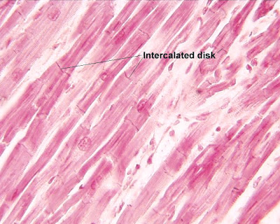
When you view cardiac muscle with a microscope a particularly striking feature becomes apparent the muscle cells exhibit a characteristic branching pattern. Notice in the pictures on this page that the parallel muscle cells are interconnected by diverging branches. Furthermore, one or sometimes two nuclei are present within each cell and are more centrally located than the nuclei of skeletal muscle cells.


The image on the left shows a photomicrograph of cardiac muscle through a light microscope.

Another unique feature of cardiac muscle is the presence of dense bands called intercalated discs that separate individual cells from one another at their ends. These discs represent specialised cell junctions between cardiac muscle cells.

These junctions offer very little resistance to the passage of an action potential from one cell to the next. The resistance here is so low that ions move freely through this permeable junction and thus permit the entire atrial or ventricular muscle mass to function as one giant cell. For this reason cardiac muscle is frequently referred to as a functional syncytium, a single functional unit.

Because cardiac muscle functions as a syncytium, stimulation of an individual muscle cell results in the contraction of all the muscle cells. This is an application of the all-or-nothing principle. Although the principle applies only to individual cells in skeletal muscle, if the stimulus in cardiac muscle is great enough to initiate contraction of a single cell, the entire muscular syncytium will undergo contraction.

Due to differences between the way in which the action potential travels through cardiac muscle it contracts at a slower rate than does skeletal muscle.

The fibrocollagenous skeleton

The heart has a fibrocollagenous skeleton, the main component being the central fibrous body, located at the level of the cardiac valves.


This image shows an electron microscope view of the fibrocollagenous material which makes up the 'skeleton' of the heart. It is important not only from the point of enabling the heart to maintain it's shape but also because it prevents atrial contraction of heart muscle being automatically transferred to the ventricles unless it is triggered by the AV node.

Extensions of the central fibrous body surround the heart valves to form the valve rings which support the base of each valve. The valve rings on the left side of the heart surround the mitral and aortic valves and are thicker than those on the right side, which surround the tricuspid and pulmonary valves.

A downward extension of the fibrocollagenous tissue of the aortic valve ring forms a fibrous septum between the right and left ventricles called the membranous interventricular septum. This is a minor component of the septum between the right and left ventricles, most of which is composed of cardiac muscle covered on both sides by endocardium. It is important in that it provides for attachment of cardiac muscle and lends support to the AV valves.The membranous part is located high in the septal wall beneath the aortic valve.

The fibrocollagenous skeleton of the heart separates the atrial syncytium from the ventricular syncytium; therefore an impulse from the former must pass through specialised tissue called the AV node before triggering the latter. The connective tissue network of the fibrous skeleton lies within the septa between the atria and ventricles.

Wednesday, October 22, 2008

Coronary Circulation

By: Dr.Norzaher


The heart muscle, like every other organ or tissue in your body, needs oxygen-rich blood to survive. Blood is supplied to the heart by its own vascular system, called coronary circulation.

The aorta (the main blood supplier to the body) branches off into two main coronary blood vessels (also called arteries). These coronary arteries branch off into smaller arteries, which supply oxygen-rich blood to the entire heart muscle.

The right coronary artery supplies blood mainly to the right side of the heart. The right side of the heart is smaller because it pumps blood only to the lungs.

The left coronary artery, which branches into the left anterior descending artery and the circumflex artery, supplies blood to the left side of the heart. The left side of the heart is larger and more muscular because it pumps blood to the rest of the body.

Anatomy of Heart

By: Dr.Norzaher


Coronary Arteries
Because the heart is composed primarily of cardiac muscle tissue that continuously contracts and relaxes, it must have a constant supply of oxygen and nutrients. The coronary arteries are the network of blood vessels that carry oxygen- and nutrient-rich blood to the cardiac muscle tissue.

The blood leaving the left ventricle exits through the aorta, the body’s main artery. Two coronary arteries, referred to as the "left" and "right" coronary arteries, emerge from the beginning of the aorta, near the top of the heart.

The initial segment of the left coronary artery is called the left main coronary. This blood vessel is approximately the width of a soda straw and is less than an inch long. It branches into two slightly smaller arteries: the left anterior descending coronary artery and the left circumflex coronary artery. The left anterior descending coronary artery is embedded in the surface of the front side of the heart. The left circumflex coronary artery circles around the left side of the heart and is embedded in the surface of the back of the heart.

Just like branches on a tree, the coronary arteries branch into progressively smaller vessels. The larger vessels travel along the surface of the heart; however, the smaller branches penetrate the heart muscle. The smallest branches, called capillaries, are so narrow that the red blood cells must travel in single file. In the capillaries, the red blood cells provide oxygen and nutrients to the cardiac muscle tissue and bond with carbon dioxide and other metabolic waste products, taking them away from the heart for disposal through the lungs, kidneys and liver.

When cholesterol plaque accumulates to the point of blocking the flow of blood through a coronary artery, the cardiac muscle tissue fed by the coronary artery beyond the point of the blockage is deprived of oxygen and nutrients. This area of cardiac muscle tissue ceases to function properly. The condition when a coronary artery becomes blocked causing damage to the cardiac muscle tissue it serves is called a myocardial infarction or heart attack.

Superior Vena Cava
The superior vena cava is one of the two main veins bringing de-oxygenated blood from the body to the heart. Veins from the head and upper body feed into the superior vena cava, which empties into the right atrium of the heart.

Inferior Vena Cava
The inferior vena cava is one of the two main veins bringing de-oxygenated blood from the body to the heart. Veins from the legs and lower torso feed into the inferior vena cava, which empties into the right atrium of the heart.

Aorta
The aorta is the largest single blood vessel in the body. It is approximately the diameter of your thumb. This vessel carries oxygen-rich blood from the left ventricle to the various parts of the body.

Pulmonary Artery
The pulmonary artery is the vessel transporting de-oxygenated blood from the right ventricle to the lungs. A common misconception is that all arteries carry oxygen-rich blood. It is more appropriate to classify arteries as vessels carrying blood away from the heart.

Pulmonary Vein
The pulmonary vein is the vessel transporting oxygen-rich blood from the lungs to the left atrium. A common misconception is that all veins carry de-oxygenated blood. It is more appropriate to classify veins as vessels carrying blood to the heart.

Right Atrium
The right atrium receives de-oxygenated blood from the body through the superior vena cava (head and upper body) and inferior vena cava (legs and lower torso). The sinoatrial node sends an impulse that causes the cardiac muscle tissue of the atrium to contract in a coordinated, wave-like manner. The tricuspid valve, which separates the right atrium from the right ventricle, opens to allow the de-oxygenated blood collected in the right atrium to flow into the right ventricle.

Right Ventricle
The right ventricle receives de-oxygenated blood as the right atrium contracts. The pulmonary valve leading into the pulmonary artery is closed, allowing the ventricle to fill with blood. Once the ventricles are full, they contract. As the right ventricle contracts, the tricuspid valve closes and the pulmonary valve opens. The closure of the tricuspid valve prevents blood from backing into the right atrium and the opening of the pulmonary valve allows the blood to flow into the pulmonary artery toward the lungs.

Left Atrium
The left atrium receives oxygenated blood from the lungs through the pulmonary vein. As the contraction triggered by the sinoatrial node progresses through the atria, the blood passes through the mitral valve into the left ventricle.

Left Ventricle
The left ventricle receives oxygenated blood as the left atrium contracts. The blood passes through the mitral valve into the left ventricle. The aortic valve leading into the aorta is closed, allowing the ventricle to fill with blood. Once the ventricles are full, they contract. As the left ventricle contracts, the mitral valve closes and the aortic valve opens. The closure of the mitral valve prevents blood from backing into the left atrium and the opening of the aortic valve allows the blood to flow into the aorta and flow throughout the body.

Papillary Muscles
The papillary muscles attach to the lower portion of the interior wall of the ventricles. They connect to the chordae tendineae, which attach to the tricuspid valve in the right ventricle and the mitral valve in the left ventricle. The contraction of the papillary muscles opens these valves. When the papillary muscles relax, the valves close.

Chordae Tendineae
The chordae tendineae are tendons linking the papillary muscles to the tricuspid valve in the right ventricle and the mitral valve in the left ventricle. As the papillary muscles contract and relax, the chordae tendineae transmit the resulting increase and decrease in tension to the respective valves, causing them to open and close. The chordae tendineae are string-like in appearance and are sometimes referred to as "heart strings."

Tricuspid Valve
The tricuspid valve separates the right atrium from the right ventricle. It opens to allow the de-oxygenated blood collected in the right atrium to flow into the right ventricle. It closes as the right ventricle contracts, preventing blood from returning to the right atrium; thereby, forcing it to exit through the pulmonary valve into the pulmonary artery.

Mitral Value
The mitral valve separates the left atrium from the left ventricle. It opens to allow the oxygenated blood collected in the left atrium to flow into the left ventricle. It closes as the left ventricle contracts, preventing blood from returning to the left atrium; thereby, forcing it to exit through the aortic valve into the aorta.

Pulmonary Valve
The pulmonary valve separates the right ventricle from the pulmonary artery. As the ventricles contract, it opens to allow the de-oxygenated blood collected in the right ventricle to flow to the lungs. It closes as the ventricles relax, preventing blood from returning to the heart.

Aortic Valve
The aortic valve separates the left ventricle from the aorta. As the ventricles contract, it opens to allow the oxygenated blood collected in the left ventricle to flow throughout the body. It closes as the ventricles relax, preventing blood from returning to the heart.


Introduction

By: Dr.Norzaher


The cardiovascular/circulatory system transports food, hormones, metabolic wastes, and gases (Oxygen, carbon dioxide) to and from cells all over the body.

Components of the circulatory system include:
blood: consisting of liquid plasma and cells
blood vessels (vascular system): the "channels" (arteries, veins, capillaries) which carry blood to/from all tissues. (Arteries carry blood away from the heart. Veins return blood to the heart. Capillaries are thin-walled blood vessels in which gas/ nutrient/ waste exchange occurs.)
heart: a muscular pump to move the blood

There are two circulatory "circuits":
Pulmonary circulation, involving the "right heart," delivers blood to and from the lungs. The pulmonary artery carries oxygen-poor blood from the "right heart" to the lungs, where oxygenation and carbon-dioxide removal occur. Pulmonary veins carry oxygen-rich blood from tbe lungs back to the "left heart."
Systemic circulation, driven by the "left heart," carries blood to the rest of the body. Food products enter the sytem from the digestive organs into the portal vein. Waste products are removed by the liver and kidneys. All systems ultimately return to the "right heart" via the inferior and superior vena cavae.

A specialized component of the circulatory system is the lymphatic system, consisting of a moving fluid (lymph/interstitial fluid); vessels (lymphatics); lymph nodes, and organs (bone marrow, liver, spleen, thymus). Through the flow of blood in and out of arteries, and into the veins, and through the lymph nodes and into the lymph, the body is able to eliminate the products of cellular breakdown and bacterial invasion.