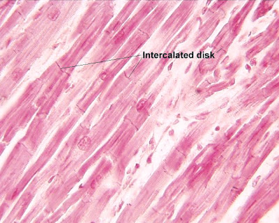
The image below shows a section through the wall of the heart in the middle of the right ventricle. Note the branched appearance of the cardiac muscle in the myocardium.
The pericardium
Within the mediastinum, the heart and origins of the great blood vessels are enclosed in a loose fitting sac termed the pericardium. This protective sac is composed of two layers separated by a space called the pericardial cavity. The outer layer is termed the parietal pericardium, and the inner layer is called the visceral pericardium.
The parietal pericardium consists of an outer layer of thick, fibrous connective tissue and an inner serous layer. The serous layer, consisting largely of mesothelium together with a small amount of connective tissue, forms a simple squamous epithelium and secretes a small amount of fluid. Normally the total volume of this fluid is only about 25 to 35 ml.
This fluid layer lubricates the surfaces to allow friction free movement of the heart within the pericardium during it's muscular contractions.
The fibrous layer of the parietal pericardium is attached to the diaphragm and fuses with the outer wall of the great blood vessels entering and leaving the heart. Thus, the parietal pericardium forms a strong protective sac for the heart and serves also to anchor it within the mediastinum.
The visceral pericardium is also known as the epicardium and as such comprises the outermost layer of the heart proper.
The epicardium (or visceral pericardium) forms the outer covering of the heart and has an external layer of flat mesothelial cells. These cells lie on a stroma of fibrocollagenous support tissue, which contains elastic fibres, as well as the large arteries supplying blood to the heart wall, and the larger venous tributaries carrying blood from the heart wall. The large arteries (coronary arteries) and veins are surrounded by adipose tissue, which expands the epicardium.

The image on the left is a diagrammatic representation of a section through the left and right ventricles as viewed from above.
Notice the difference in thickness between the wall of the left ventricle and that of the right ventricle. Also note their different shapes.
Atrial myocardial fibres are not only smaller than those of the ventricles but also contain small neuroendocrine granules, which are usually sparse and located close to the nucleus; they are most numerous in the right atrium. These granules secrete atrial natriuretic hormone when the atrial fibres are stretched excessively.
Atrial natriuretic hormone increases the excretion of water and sodium and potassium ions by the distal convoluted tubule of the kidney. It also decreases blood pressure by inhibiting renin secretion by the kidneys and aldosterone secretion by the adrenals.
The right ventricle pushes blood through the pulmonary valve and through the lungs, to enter the left atrium. It therefore has a moderately thick muscle layer composed of fibres intermediate in diameter between atrial and left ventricular muscle cells.
The left ventricle pumps blood through the high-pressure systemic arterial system and therefore has the thickest myocardium with the largest diameter muscle fibres.
In both ventricles, raised mounds of cardiac muscle (papillary muscles) protrude into the ventricular lumina and point toward the atrioventricular valves. Papillary muscles are the site of attachment of chordae tendinae, narrow tendinous cords that tether the valve parts to the wall of the ventricle beneath them.
The endocardium
The internal lining of all four heart chambers is the endocardium, which is composed of three layers
| a layer in direct contact with the myocardium | |
| a middle layer | |
| an innermost layer. |
The outmost layer is composed of irregularly arranged collagen fibres that merge with collagen surrounding adjacent cardiac muscle fibres. This layer may contain some Purkinje fibres, which are part of the impulse conducting system as we shall see later.
The middle layer is the thickest endocardial layer and is composed of more regularly arranged collagen fibres containing variable numbers of elastic fibres, which are compact and arranged in parallel in the deepest part of the layer. Occasional myofibroblasts are present.
The inner layer is composed of flat endothelial cells, which are continuous with the endothelial cells lining the vessels entering and emerging from the heart.
The endocardium is variable in thickness, being thickest in the atria and thinnest in the ventricles, particularly the left ventricle. The increased thickness is due almost entirely to a thicker fibroelastic middle layer. Localised areas of endocardial thickening (jet lesions) are common, particularly in the atria, and result from turbulent blood flow within the chamber.
Cardiac muscle
Cardiac muscle and skeletal muscle have some features in common. Like skeletal muscle, cardiac muscle is striated, has dark Z lines, and possesses myofibrils that contain actin and myosin filaments. During systole these filaments slide over one another in much the same manner as in skeletal muscle contraction.
On the other hand, cardiac muscle has several unique properties. It is capable of intrinsic contraction (contraction without being triggered by a nerve impulse). This is a characteristic of all cardiac muscle. Skeletal muscle, however, contracts only when stimulated via a nerve impulse.

When you view cardiac muscle with a microscope a particularly striking feature becomes apparent the muscle cells exhibit a characteristic branching pattern. Notice in the pictures on this page that the parallel muscle cells are interconnected by diverging branches. Furthermore, one or sometimes two nuclei are present within each cell and are more centrally located than the nuclei of skeletal muscle cells.
The image on the left shows a photomicrograph of cardiac muscle through a light microscope.
Another unique feature of cardiac muscle is the presence of dense bands called intercalated discs that separate individual cells from one another at their ends. These discs represent specialised cell junctions between cardiac muscle cells.
These junctions offer very little resistance to the passage of an action potential from one cell to the next. The resistance here is so low that ions move freely through this permeable junction and thus permit the entire atrial or ventricular muscle mass to function as one giant cell. For this reason cardiac muscle is frequently referred to as a functional syncytium, a single functional unit.
Because cardiac muscle functions as a syncytium, stimulation of an individual muscle cell results in the contraction of all the muscle cells. This is an application of the all-or-nothing principle. Although the principle applies only to individual cells in skeletal muscle, if the stimulus in cardiac muscle is great enough to initiate contraction of a single cell, the entire muscular syncytium will undergo contraction.
Due to differences between the way in which the action potential travels through cardiac muscle it contracts at a slower rate than does skeletal muscle.
The fibrocollagenous skeleton
The heart has a fibrocollagenous skeleton, the main component being the central fibrous body, located at the level of the cardiac valves.
This image shows an electron microscope view of the fibrocollagenous material which makes up the 'skeleton' of the heart. It is important not only from the point of enabling the heart to maintain it's shape but also because it prevents atrial contraction of heart muscle being automatically transferred to the ventricles unless it is triggered by the AV node.
Extensions of the central fibrous body surround the heart valves to form the valve rings which support the base of each valve. The valve rings on the left side of the heart surround the mitral and aortic valves and are thicker than those on the right side, which surround the tricuspid and pulmonary valves.
A downward extension of the fibrocollagenous tissue of the aortic valve ring forms a fibrous septum between the right and left ventricles called the membranous interventricular septum. This is a minor component of the septum between the right and left ventricles, most of which is composed of cardiac muscle covered on both sides by endocardium. It is important in that it provides for attachment of cardiac muscle and lends support to the AV valves.The membranous part is located high in the septal wall beneath the aortic valve.
The fibrocollagenous skeleton of the heart separates the atrial syncytium from the ventricular syncytium; therefore an impulse from the former must pass through specialised tissue called the AV node before triggering the latter. The connective tissue network of the fibrous skeleton lies within the septa between the atria and ventricles.


No comments:
Post a Comment